.
Organoids offer a window into early brain development that's impossible to observe closely in humans, namely because it unfolds inside developing fetuses. Although organoids are not perfect re-creations of full-size human brains, scientists think organoids are useful models for studying the developing brain in both health and disease.
The model in the new study "certainly creates some pretty interesting options in terms of modeling human neurodevelopment and various types of neurodevelopmental disorders," Chen said. "If you're looking for a model that really allows you to look at larger areas of human neural tissue from a cellular, molecular perspective, I think there's a lot that this model has to offer."
 S. Pasca lab, Stanford University
 S. Pasca lab, Stanford University
 S. Pasca lab, Stanford University
Making space for human cells
Often, brain organoids are grown outside living organisms, either in lab dishes or devices that keep these "minibrains" suspended in a solution. Multiple organoids representing different parts of the brain, or even different people's brains, can also be brought together to form more-complex structures.
Why, then, are some scientists growing human organoids inside mice? One reason is that there's a secret sauce inside living organisms that helps organoids mature better in animals than they do in lab dishes. In the body (in vivo), there are mysterious signals that help direct neurons' development and organization, and these signals are missing in lab dishes (in vitro).
"There are some cues that are present in vivo that are really important, and we simply don't know what to add in, in vitro," said study co-author Dr. Sergiu Pașca, a professor of psychiatry and behavioral sciences at Stanford University. The team demonstrated this phenomenon by transplanting organoids into lab rats in a study published in 2022; the transplanted organoids grew larger, formed better connections and were more active than organoids grown in dishes.
But transplanting human brain cells into rodents comes with different challenges. One is that human brains mature at a slower rate than rodent brains do. "Even when they're put in an animal, in a mouse or in a rat, they will still develop about 20 times slower than the mouse or the rat," Pașca told Live Science.
The host's brain cells quickly grow and form new connections, while the human cells lag behind and get outcompeted, Pașca explained. This limits the amount of space that the human cells can take up. As neurons mature, they become myelinated, meaning they gain fatty insulation that helps them communicate more efficiently. The rodent neurons become myelinated more quickly than the human cells do, and that fat creates a physical barrier that the human cells struggle to penetrate, Pașca said.
In their study, described Wednesday (Sept. 16) in the journal Nature, Pașca and colleagues aimed to give the human neurons a leg up. They couldn't solve the issue of human cells maturing slowly, but they could lend the cells extra space to grow in.
Through years of work, they developed a genetically modified mouse that develops only 2% of its cerebral cortex. The majority of its hippocampus — a major memory center in the brain — is also missing. Within a few days of the mouse's birth, the team transplants human neural tissue into that vacant space, and about 90% of the time, that human tissue integrates successfully and starts to grow, Pașca said.
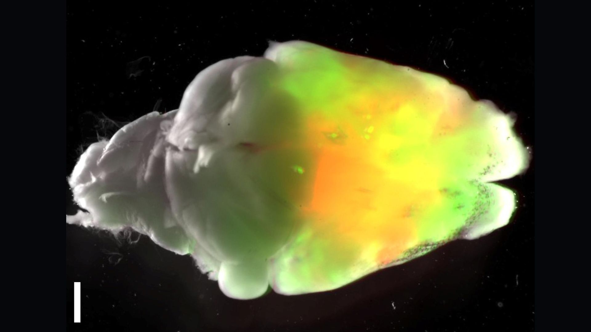
The brains of normal lab mice look like the brain on the left, whereas the center brain is missing 98% of its cortex and hippocampus. The brain on the right has human cortical tissue added to it. (Here, "XCX" is short for "xenocortical," and "xeno" references the transfer of tissues from one species to another.) (Image credit: Pasca Lab/Stanford University)"We just took cortical organoids, about four of them, and transferred them with a syringe into that vacant space," he said. "That's it. They go in there, they graft, and within a few weeks, they start to grow. And then within a few months, they've taken most of that volume."
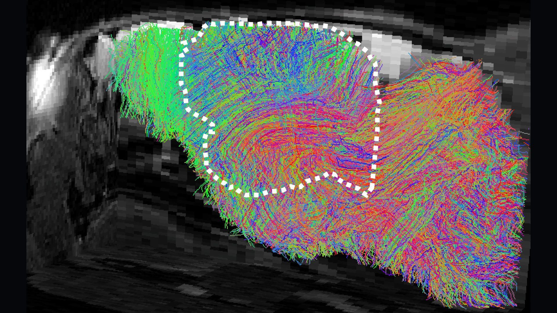
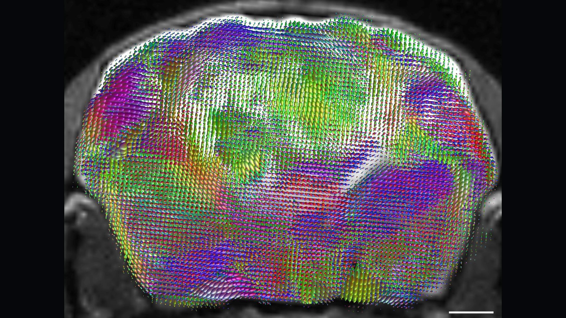
In the weeks following the transplantation procedure, the human brain cells in the mice's heads grew, formed connections and extended projections into the underlying mouse tissue. The human tissue didn't organize itself into distinct layers as it normally would inside a human's head, but it included many cell types that are typically seen in the human cerebral cortex.
Future of the field
The researchers compared the mice imbued with brain organoids with mice that were missing the same amount of brain tissue but didn't get organoids. They also compared both groups to unmodified lab mice.
Perhaps surprisingly, the mice missing large chunks of their brains still functioned fairly well. "You look at them, and you can't really honestly tell," Pașca said. Upon closer inspection, though, the mice had subtle deficits in their fine-motor skills, working memory and socialization, he noted.
"From our perspective as humans, so much of what we do day to day depends on the cortex," Chen noted. But in a mouse, the cortex makes up a minority of the overall brain, and various studies have suggested that the animals can get by without it, he said. "I don't think the transplantation process itself is significantly hurting the animal," he added.
It would be much more problematic if this was to happen in a species that has a larger brain and is closer to humans evolutionarily.
Dr. Sergiu Pașca, a professor of psychiatry and behavioral sciences at Stanford University
In the future, the ability to compare these three sets of mice — with organoids, without organoids, and unmodified — could help scientists tease apart how the human tissue is contributing to a given experimental result. Pașca envisions that the approach will be useful for studying the effects of insults to the brain, such as hypoxia (low oxygen) and exposure to toxins or drugs in the womb. Additionally, the genetics of the human organoids could be tweaked to see how those changes affect the brain's development, structure and function. That could be useful for studying cerebral palsy or autism, he suggested.
Chen expects that this new approach will be useful for studying aspects of early brain development at the molecular and cellular levels. The new model incorporates a larger volume of human tissue than models have done in the past, and that's an additional advantage. The human tissue doesn't organize itself into layers or lobes as you'd see in a real human brain, he noted, but he thinks there's still a lot that can be learned from it.
From an ethical standpoint, Pașca consulted with experts at Stanford and an external ethics committee regarding the welfare of the animals used in the study. Studies that involve putting human brain tissue into animals also raise questions about whether that added tissue could grant the animals new cognitive abilities — make them more human-like, in essence. The ethics committee fielded these concerns as well.
"We have not seen emergence of any new properties," Pașca noted. "It's not surprising because we're still at a very early stage of development." They grew the organoids for only six months, so the human organoids were roughly as developed as a 6-month-old fetus's brain tissue.
If the organoids matured to be more human-like — developing layers and lobes — that might present more of a concern. But both Pașca and Chen said that type of development might be hard to recreate in a mouse anyway.
Chen argued that the sheer size of the human brain contributes to its complexity, and it also contains specialized regions that work together to execute different tasks. A mouse's head cannot support the scale of a human brain, and at this point, organoids don't develop the same organization and specialization that we see in people, he said.
"It would be much more problematic if this was to happen in a species that has a larger brain and is closer to humans evolutionarily," Pașca argued. Pigs and nonhuman primates, such as monkeys, would be examples. Especially in regard to transplanting human organoids into monkeys, "that would be an experiment that I don't see is justified at this point," Pașca said.
See how much you know about the most complex organ in the human body with our brain quiz!
'> Scientists shrank mice's brains and replaced the missing tissue with human 'organoids'
"It is extremely rare to witness," study first author Olaf Meynecke, a marine ecologist at Griffith University in Australia, told Live Science in an email. "Only about 20 births of humpback whales have been observed to date and described in literature," none of which involved a stillborn calf.
Unlike the babies of toothed whales and porpoises, newborn humpback whales don't have enough blubber to float, so they sink without the help of an adult whale. This might partly explain why scientists had not witnessed a stillbirth in this species until now, Meynecke said, as the lifeless body sinks to the seabed immediately after birth and is almost impossible to spot from a distance.
The whale mom repeatedly tried to make eye contact with her calf, even though researchers think she was aware it died during birth. (Image credit: Andy Mulville)The video was taken after study co-author Andrew Mulville, who was on a private boat that morning, noticed two adult humpback whales milling about at the surface of the water. One of the whales adopted a vertical position with its tail pointing up. Moments later, Mulville saw the remains of an amniotic membrane, the inner layer of the placental bag that envelops a fetus, floating on the water.
The whale had obviously just given birth, but there was no sign of the calf. So Mulville and others on the boat followed the female underwater with a GoPro camera. They found the calf dead on the seafloor, while the mother displayed what the study authors interpreted as "postmortem attentive behavior," or grieving.
"She was repeatedly diving down for short times and swimming next to the calf, then resting next to it on the floor for about a minute, even moving the pectoral fin over the calf, then diving up again," Meynecke said. This behavior lasted for at least 90 minutes and possibly days, although the researchers left the scene after about two hours of observation.
The mother did not try to bring the calf's body to the surface, possibly because she realized that it had died during birth. This is different from the postmortem attentive behavior of toothed whales, which researchers have previously seen pushing stillborn babies above the water, according to the study.
The other adult whale on the scene was an "escort" supporting the pregnant female as she prepared to give birth, Meynecke said. Based on the escort's size and appearance, it was likely a female of a similar age to the mom, but the researchers did not confirm this. Male and female escorts are common in humpback whales; they usually travel with females and their calves during migration and assist females during pregnancy.
Grieving behavior suggests that humpback whales have an inherent, strong maternal attachment to their calves. Grief is difficult to demonstrate due to the lack of physiological or neurological measurements. However, the whale mom sought close proximity with her calf during the observation time, and the adult whales were spotted swimming in the area for the next two days, suggesting they were emotionally invested in the death, according to the study.
"It provides a rare window into the behavioural response of baleen [filter-feeding] whales toward a deceased member of their species but also in this case of a mother toward her calf," Meynecke said. "It provides important insight into the complex behaviours of the species and aligns with other observations of care taking, assisting distressed whales or even protecting other species from predators."
'> 'It is extremely rare to witness': Scientists record humpback whale mom grieving her stillborn calf in first-of-its-kind footage What happened next: Because the man had fainted twice and had a history of heart disease, doctors first looked for problems affecting his brain and heart. An MRI of his brain, used to look for structural abnormalities that could explain the fainting, and an electroencephalogram (EEG), which records the brain's electrical activity, did not reveal a cause.
The man had persistently low blood pressure, the doctors observed, and this led them to move him to the intensive care unit. There, doctors carried out tests for infections and heart attack, but all came back negative.
Meanwhile, the swelling around one of his eyes spread to both eyes and then across his face, while his blood pressure continued to fall despite treatment with intravenous fluids.
The doctors probed further into the man's medical history and activities before the fainting episodes. They learned that he had applied an artificial hair dye two days before he became ill. Soon afterward, he developed itching on his scalp, neck and face. He self-treated with the allergy medicine cetirizine, an antihistamine, for two days. He also reported spending two to three hours outdoors in the sun each day after he'd applied the hair dye.
The diagnosis: Based on the man's recent use of hair dye, the itching that followed, his rapidly worsening facial swelling and his persistently low blood pressure, doctors concluded that he was experiencing anaphylaxis, a severe allergic reaction that can affect the whole body. The reaction can be life-threatening if left untreated.
Anaphylaxis is a medical emergency and can be life-threatening. (Image credit: SCIENCE PHOTO LIBRARY via Getty Images)The treatment: Doctors treated the man with epinephrine (also called adrenaline), a hormonal medication that rapidly reverses the effects of anaphylaxis. They also gave him antihistamines and steroids to help bring the allergic reaction under control, along with intravenous fluids to raise his blood pressure.
However, the patient's blood pressure remained dangerously low, so doctors continued giving him epinephrine through an intravenous drip for about 16 hours. Over the next three days of treatment, his blood pressure stabilized and the swelling in his face gradually subsided.
He was transferred to a general ward and treated there for another five days before being discharged. He was advised that he could have a similar reaction in the future and referred to a dermatologist.
What makes the case unique: Most allergic reactions to hair dye remain limited to the skin, causing itching, redness and swelling where the dye has touched it. Severe reactions affecting the whole body are uncommon, the man's doctors wrote in a report of the case.
The itching the man noticed shortly after applying the dye was likely an early, mild allergic reaction, his doctors wrote. That reaction later progressed to a much more severe one, they suggested, raising the possibility of a "biphasic" allergic reaction, an uncommon pattern seen in about 5% of patients with anaphylaxis. However, this remains only a possible explanation.
The doctors also noted that the man's prolonged sun exposure after he applied the dye may have intensified the reaction; they cited previous research that ultraviolet light can enhance the allergy-triggering effects of para-phenylenediamine, a common ingredient in hair dyes. In the report, the doctors did not identify the exact hair dye ingredient that triggered the reaction in this instance.
The case highlights the importance of asking about recent use of products such as hair dye when doctors evaluate patients with otherwise unexplained fainting, facial swelling or shock, the medical team concluded.
For more intriguing medical cases, check out our Diagnostic Dilemma archives.
This article is for informational purposes only and is not meant to offer medical advice.
Can you guess the diagnosis in these strange medical cases? Find out with our diagnostic dilemma quiz!
'> Diagnostic dilemma: A man's sudden fainting episodes landed him in the hospital — and a hair dye was to blame Lenovo representatives unveiled their new AeroBlade machine at Lenovo Innovation World '26, which took place Sept. 3 at IFA 2026 in Berlin, which itself ran between Sept. 4 and 8.
Made by engineers at Frore Systems, the low-power AirJet Mini chip measures 1.63 x 1.06 x 0.1 inches (41.5 x 27 x 2.65 mm) and weighs just 0.02 pounds (9 grams). Because it contains tech designed to replace conventional cooling technologies, the AeroBlade, which is fitted with a 14-inch (355.6 mm) OLED display, is much thinner and lighter than comparable laptops
The AeroBlade measures less than 0.39 inches (10 mm) thick — where comparably sized machines measure 0.51 inches (12 mm) at their thinnest — and weighs just 1.8 pounds (830 g). Laptops as light as this exist, like the 1.76-pound (799 g) Acer Swift Blade 14, but they're rare. Counterparts with 14-inch screens are normally considered light if they fall between 2.2 to 2.86 pounds (1 to 1.3 kilograms).
A new approach to keeping cool
Four AirJet Mini chips are fitted inside the new Lenovo concept laptop. (Image credit: Lenovo and Frore Systems)Both companies are framing the introduction of this technology as a way to cool down the next generation of high-end laptops that will wield on-device artificial intelligence (AI).
Most laptops marketed as "AI PCs" just feature a neural processing unit (NPU) that can run simple AI workloads, such as manipulating or enhancing a webcam's video feed in real time.
But some future laptops will aim to let users run powerful large language models or AI agents without needing to connect to the internet. An example is the forthcoming series of Nvidia RTX Spark laptops, which will feature a new chip designed specifically to orchestrate AI agents locally.
Running on-device AI models usually demands significant power funneled into professional-grade graphics processing units (GPUs). Fans are deployed in all kinds of laptops to keep their chips cool, but they can be noisy. Instead of or in addition to fans, passive heat sinks, found in machines like the MacBook Air or the Microsoft Surface Pro, use metal plates positioned close to heat sources to absorb thermal energy. But these laptops are more prone to throttling — when their software pulls back performance to reduce temperature.
The AirJet Mini is a different way of cooling, relying on its own high-velocity jet mechanism to flush hot air away. Frore representatives said in a statement that the device operates at just 21 decibels at peak power — equivalent to the sound of leaves rustling or a whisper — and is capable of removing 7.5 watts of heat. Four of these chips would remove 30W of heat — which is in line with the heat expulsion of conventional laptop fans, although there is variation between models.
The AeroBlade's 2,000 pascals of back pressure a measure of opposing force pushing against the direction of airflow) is roughly 10 times that found in regular laptop fans, which typically falls between 100 and 200 pascals.
Although the underlying cooling technology is a few years old, having first been announced in December 2022, AeroBlade represents the first time it's been used in a fully operational laptop. The chip was previously integrated into the ZOTAC ZBOX PI430AJ Pico mini PC in 2023, while there are no commercially available tablets or laptops available with this chip. The AeroBlade laptop remains a concept model, with no information yet as to when production-ready machines fitted with this new cooling technology will emerge.
Can you match these ancient devices to their pictures? Find out with our computing quiz!
'> New laptop ditches noisy fans and heat sinks for tiny 'cooling chips' — it's much thinner and lighter than you'd expect Science Advances.
"By comparing JWST observations with those obtained … over the last decade, we discovered opposite changes in the two rings: while the inner ring shows significantly higher opacity, the outer ring shows lower opacity, " Pablo Santos-Sanz, a researcher at the Institute of Astrophysics of Andalusia and first author of the study, said in a statement.
Researchers don't know exactly how the changes took place, but the study authors offered three suggestions. The first is that JWST, which has a fine resolution, was able to see both denser and sparser zones of the rings than previous observations showed. The second is that there were changes in the rings' material or in the grains themselves. Finally, a third possibility is that JWST could see "grains with wavelength-dependent optical properties," meaning the telescope saw effects in composition and grain size that explain the variability.
Santos-Sanz said the research suggests that small bodies with rings — even those orbiting far away from the gravitational influences of planets — may have more changes in their systems than scientists previously thought. "Our results force us to rethink how they form, how they evolve, and what mechanisms maintain their stability," he said.
A theoretical follow-up observation, taken the next time Chariklo passes in front of a star, would help to confirm the new JWST results, the researchers added — especially the apparent decreased opacity of one of the rings and the increased opacity of the other.
"These observations would help disentangle temporal evolution from wavelength-dependent scattering effects, and clarify the physical processes shaping Chariklo’s rings," the researchers stated in the study.
A JWST milestone
The new observations of Chariklo also mark the first time that scientists have planned a JWST viewing campaign specifically around a stellar occultation — that is, the researchers timed the viewing precisely to when Chariklo passed in front of a distant star from the telescope's point of view.
All observations of Chariklo's rings, on the ground and in space, have used stellar occultations. That's because the rings are too faint to be imaged directly, so astronomers must instead learn more about the rings by measuring the dips and increases in brightness as Chariklo passes in front of the background star. Scientists use a similar method to study the atmospheres of alien planets with JWST's infrared instruments.
Chariklo's speed relative to JWST was low, at 1.5 miles per second (2.5 kilometers per second), allowing for fine imaging of the rings. To time the occultation, the researchers used info from the European Space Agency's Gaia mission, which precisely charts the locations and movements of more than 2 billion stars, as well as calculated the orbit of JWST at a location roughly 930,000 miles (1.5 million km) from Earth in the opposite direction of the sun. According to the researchers, the planning and execution of the mission required "extreme precision."
'> Strange 'centaur' object between Saturn and Uranus has mysterious, changing rings, James Webb telescope study hints